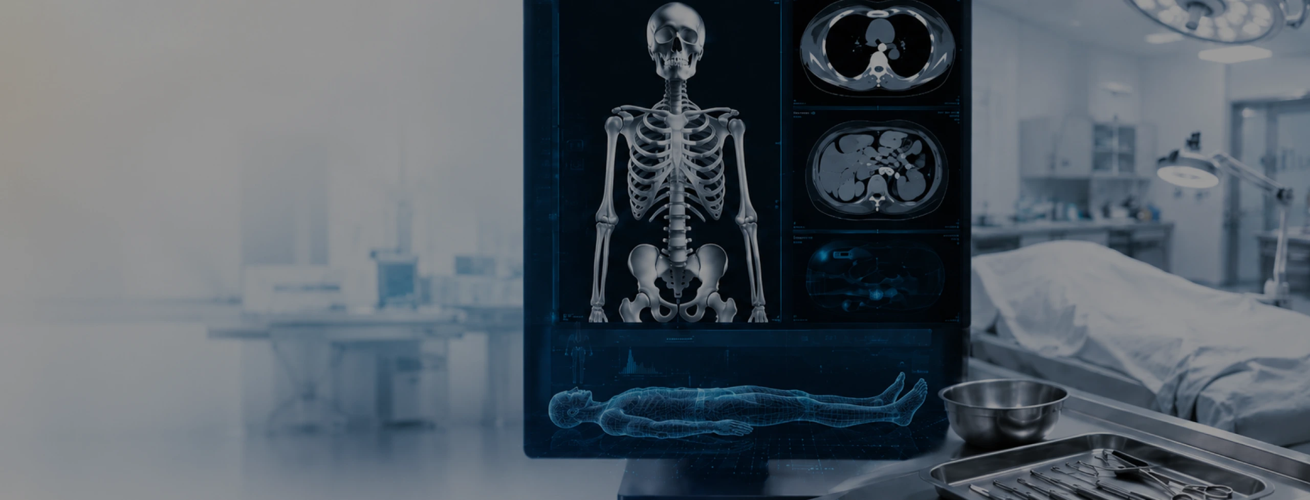
Virtual Autopsy vs Conventional Autopsy: Understanding Their Complementary Roles
Summary
Virtual autopsy and conventional autopsy offer different ways of examining evidence after death. Understanding where each method's strengths lie can help forensic teams build more complete investigations while improving documentation, reviewability and long-term learning.
What can one method see that the other may miss?
When people discuss virtual autopsy in forensic investigation, the conversation often turns into a comparison with conventional autopsy. Which is better? The obvious answer: both.
These questions are understandable, but they can also narrow the discussion too quickly. For forensic teams, the more useful question is often what each method contributes to the investigation. Medical imaging can reveal and preserve findings that are difficult to appreciate through dissection alone. Conventional autopsy, meanwhile, remains essential for findings that require direct examination, tissue sampling and laboratory analysis. Understanding how those strengths fit together is where the conversation becomes more interesting.
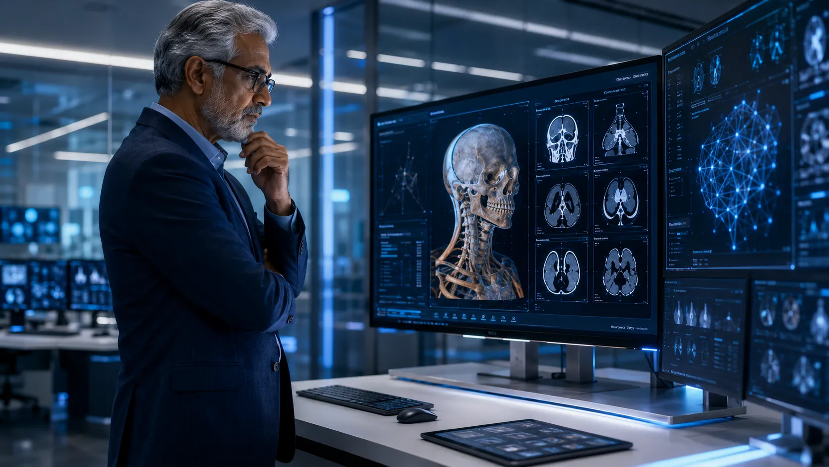
Virtual autopsy vs conventional autopsy: what does each reveal?
Conventional autopsy allows the forensic expert to directly examine organs, tissues and injuries and collect samples for histopathology, toxicology and other investigations. It remains fundamental to forensic medicine. Virtual autopsy, commonly involving post-mortem CT (PMCT) and in some settings MRI or other imaging techniques, adds another layer of information. Its strengths can be particularly useful for examining:
skeletal fractures and injuries
foreign bodies and their location
gas collections
certain haemorrhages
anatomical relationships between injuries
three-dimensional injury patterns
A systematic review comparing post-mortem imaging with conventional autopsy in trauma victims found that PMCT was particularly sensitive in detecting skeletal injuries, while conventional autopsy performed better for minor organ and soft-tissue injuries. The researchers concluded that PMCT should be considered an important supplement to conventional autopsy in trauma cases.
That distinction matters. An investigation benefits when the forensic expert can move between different forms of evidence rather than relying on a single view of the body.
Imaging can give the forensic expert a map before examination begins
One of the most practical benefits of virtual autopsy appears before an invasive examination even starts. Consider a complex trauma case involving multiple fractures, internal injury and foreign material. A whole-body scan can provide an overview of the body and help identify areas that deserve closer examination. The forensic expert therefore begins with additional information.
Earlier research into PMCT in trauma cases found that imaging could identify injuries missed during conventional examination and suggested that pre-autopsy CT could help guide pathologists towards injuries that may otherwise be difficult to locate. This creates a useful workflow:
- Capture the body digitally.
- Review the imaging findings.
- Identify areas requiring particular attention.
- Conduct the conventional examination where necessary.
- Correlate imaging, pathology and other evidence.
Forensic imaging in investigations can therefore contribute before, during and after the physical examination.

Virtual autopsy can strengthen the evidence available to forensic experts
One of the strengths of virtual autopsy is its ability to examine the body as a complete digital dataset before any invasive procedure alters it. Post-mortem CT can document skeletal injuries, gas collections, foreign bodies, major haemorrhages and anatomical relationships between findings. Research comparing PMCT with conventional autopsy has demonstrated particular strengths in areas such as fractures, gas and foreign-body detection. This gives forensic experts an additional source of evidence to examine alongside case history, laboratory findings, and, where required, further physical examination.
The value becomes even greater because the imaging remains available after the investigation. A finding can be viewed again, measured from another angle, discussed with another specialist or revisited when new questions arise.
Of course, some forensic questions still require tissue examination, toxicology, histopathology or other investigative methods. The choice should be guided by the circumstances of the case. What virtual autopsy brings to that process is a powerful additional capability: the opportunity to see, document and preserve evidence digitally before deciding what further examination may be necessary.
What happens to the evidence after the examination?
This may be one of the most important differences for institutions thinking beyond an individual case. A conventional examination records findings through reports, photographs, samples and documentation. Imaging adds something different: a three-dimensional digital record that can be revisited. Months or even years later, another specialist can review the scan. Measurements can be repeated. Particular structures can be re-examined from another perspective. New questions can be considered against the original imaging dataset. That has implications beyond individual cases.
The same appropriately governed digital evidence can contribute to:
multidisciplinary case review
forensic education and resident training
research
second opinions
retrospective analysis
institutional knowledge
This leads to a broader way of thinking about virtual autopsy infrastructure. A scan should not be viewed only as an examination performed on a particular day. It can become part of an institution's long-term forensic knowledge base.
The future may be a layered forensic investigation
From our work in forensic technology and education, we believe the strongest model will bring different forms of evidence together. Imaging provides one layer. Physical examination provides another. Histopathology, toxicology, case history, investigative information and specialist interpretation add further layers. Technology makes those layers easier to capture, correlate, preserve, and revisit.
This thinking also shapes SEVA Technologies' approach to modern forensic infrastructure. We see virtual autopsy as part of a wider ecosystem involving medical imaging, digital workflows, education and research rather than as an isolated technology. As institutions modernise, the opportunity is to build systems in which evidence moves more effectively between investigation, interpretation, learning and future review.
Conclusion: Better forensic investigations can use the strengths of both
The discussion around virtual autopsy in forensic investigation does not need a winner. Virtual and conventional autopsy have different strengths, and their appropriate use depends on the case, legal requirements and forensic questions involved. For institutions preparing for the future, extending readiness will increasingly mean having the expertise, infrastructure and digital workflows to use these methods together responsibly.
Exploring how virtual autopsy could fit into your institution's forensic workflow?
Explore SEVA Technologies' Virtual Autopsy ecosystem or request a discussion with our team.
FAQs
Virtual autopsy uses medical imaging techniques such as post-mortem CT and, in some settings, MRI to examine the body after death. It allows forensic experts to study internal structures, fractures, foreign bodies and other injuries while complementing conventional examination and other evidence.
Not in every case. Research shows that PMCT can be particularly effective for skeletal trauma, gas and foreign bodies, while conventional autopsy remains important for soft-tissue examination, histopathology, toxicology and laboratory investigation. The appropriate approach depends on the circumstances and questions surrounding each investigation.
Beyond non-invasive examination, a key benefit is creating a permanent digital dataset. Imaging can help document injuries, guide further examination and support multidisciplinary review. With appropriate governance, stored images may also support future second opinions, teaching and research, extending their usefulness beyond the original examination.
Imaging performed before an autopsy can provide forensic experts with an overview of injuries and internal structures. This can help identify fractures, foreign objects or areas requiring closer examination. In complex trauma cases especially, having this information beforehand can help forensic teams plan and focus the subsequent physical examination.
No. Post-mortem imaging has applications across different types of death investigation, although its diagnostic strengths and limitations vary considerably depending on the case. Trauma is particularly well studied because CT is effective for visualising skeletal injuries. Forensic experts still need to determine which combination of imaging, pathology and laboratory investigation is appropriate.
Yes. A key characteristic of digital imaging is that the dataset can be stored and revisited. Subject to appropriate evidence-management and access protocols, specialists can return to the images for review, teaching, research or second opinions without needing to recreate the original examination.
The opposite is more likely. More information creates a greater need for experienced interpretation. Post-mortem imaging contains findings and changes that require specialised professional and radiological understanding. The value of combining technology with professional judgement is best understood when technology and other investigative evidence can be interpreted together.