How CT Imaging Is Transforming Death Investigations Worldwide
Summary
Postmortem CT is giving forensic experts a new way to examine trauma, document findings, and preserve evidence digitally. Its growing use worldwide is also changing how we think about the relationship between imaging, autopsy and modern death investigation.
What If We Could See More Before an Autopsy Begins?
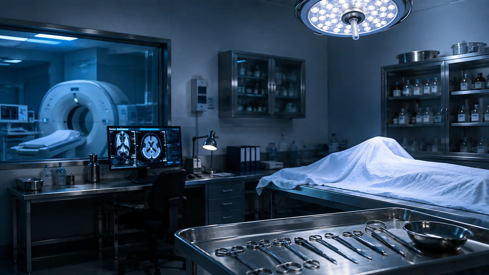
Forensic medicine has always depended on careful observation. Today, postmortem CT is adding another layer to that observation by allowing forensic experts to examine the body internally before an invasive examination begins. Fractures can be mapped, foreign bodies located, gas collections identified and findings preserved as a digital dataset.
Around the world, this is making forensic CT an increasingly important part of death investigation. But its real significance lies beyond the scan itself. It is changing what experts can see, what they can preserve, and how they can review evidence later.
Why Is Postmortem CT Gaining Ground Worldwide?
CT entered medicine as a diagnostic tool for the living. Its value after death emerged later, as forensic specialists began recognising something important: many of the qualities that make CT useful clinically become equally valuable, and sometimes uniquely useful, in a forensic examination. A 2025 review published in RadioGraphics describes PMCT as widely accepted internationally as a supplementary forensic tool and, in specific circumstances, an alternative to full autopsy.
The attraction is understandable.
A whole-body CT scan can create hundreds or thousands of images without altering the anatomy. Those images can then be reconstructed in different planes and in three dimensions. Investigators are no longer limited to what they can observe during one examination at one moment in time. That changes the possibilities considerably.
What Does CT Allow Forensic Experts to See Differently?
Postmortem CT is changing what forensic experts can observe, preserve and review. Three applications stand out.
1. Complex Trauma Can Be Viewed as a Complete Pattern
Consider a death involving extensive skeletal trauma. During an autopsy, experts can examine individual injuries carefully. With CT, forensic experts can also step back and look at their distribution across the body. Skull fractures, vertebral injuries, rib fractures and complex injuries to the pelvis can be visualised together. Three-dimensional reconstruction can make their anatomical relationships easier to understand.
This is one area where postmortem imaging has become particularly valuable. Current forensic literature consistently identifies skeletal trauma, foreign bodies and gas collections among the areas where PMCT performs strongly. For the forensic expert, the advantage is not simply finding more injuries. Seeing how injuries relate to one another may help with the larger task of reconstructing what happened.
Interested in how forensic evidence is preserved?
Explore our related article, From Crime Scene to Courtroom: The Journey of Digital Forensic Evidence.
EXPLORE MORE
2. The Body Can Be Examined Before the Anatomy Is Disturbed
Sequence matters in forensic investigation. Once an invasive examination begins, anatomy inevitably changes. CT offers an opportunity to record internal structures in situ before that happens.
locating projectiles and foreign bodies;
mapping complex fractures in situ;
recording gas collections;
guiding the subsequent examination;
preserving anatomical context before dissection.
Imagine a case involving a projectile or another foreign body. Imaging can establish its location and relationship with surrounding structures before dissection. The pathologist can then approach the subsequent examination with that information already available. The same principle applies to complex fractures and certain gas collections. This is where forensic CT works particularly well alongside conventional autopsy. Imaging provides a map. The forensic expert decides how that map should inform the rest of the investigation.
For a deeper look at what happens to forensic evidence after an examination, readers can also explore “From Crime Scene to Courtroom: The Journey of Digital Forensic Evidence.”
EXPLORE MORE
3. Evidence Can Be Reopened Without Reopening the Body
This may be one of the most important changes postmortem CT brings to forensic practice.
A conventional autopsy cannot simply be repeated in its original form. A CT dataset can be revisited. Another expert can review the images. An expert can reconsider a particular injury months later. Images can be reconstructed differently if a new question emerges. Selected findings can also be presented visually to investigators or legal professionals.
Research into PMCT visualisation has highlighted this advantage, including the usefulness of 3D representations when findings need to be communicated to people without specialist radiological knowledge. That gives forensic evidence something increasingly valuable in a digital world: a durable record that remains open to review.
Could CT Make Death Investigation More Respectful Too?
The scientific value of imaging is only one part of the conversation. Death investigation takes place within families, communities, religions and cultures. In some situations, invasive autopsy can create genuine concern because of beliefs around the treatment of the deceased. Postmortem imaging may provide another option in selected cases. International literature has documented the use of PMCT where religious or cultural concerns about invasive examination exist, although whether imaging can replace or reduce an autopsy depends on the circumstances, jurisdiction and evidence required.
India deserves particular attention. Scientific rigour and dignity do not have to be opposing ideas. As imaging capabilities develop, we can consider how to better accommodate both within modern forensic practice.
Does This Mean CT Will Replace the Conventional Autopsy?
No, and framing the conversation that way probably misses the larger opportunity. CT has limitations. Changes that are obvious during direct examination may be difficult to interpret on an unenhanced scan. Histopathology and toxicology answer questions that CT cannot. Postmortem changes can also produce imaging appearances that require specific expertise to interpret correctly. There are also areas where more specialised techniques may be required.
For example, the absence of active circulation creates challenges when examining the cardiovascular system with conventional PMCT, which is one reason postmortem CT angiography has developed. The more useful question, therefore, is: How can imaging and conventional forensic methods work together to produce a more complete investigation? That is where the future becomes interesting.
The Bigger Shift Is from a Procedure to a Forensic Ecosystem
It is tempting to think that adopting postmortem CT means installing a scanner. In practice, the scanner may be the easiest part. Useful forensic imaging requires people who understand postmortem interpretation. It needs appropriate scanning protocols, collaboration between forensic medicine and radiology, secure handling of digital evidence, reporting workflows and professionals who know when imaging findings need to be supported by autopsy, histopathology, toxicology or other investigations.
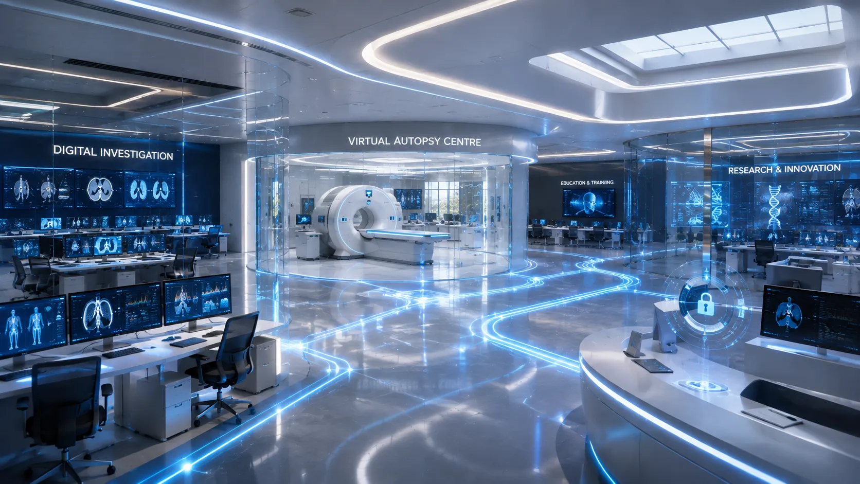
There is an educational dimension too. As forensic imaging becomes more relevant, medical colleges and forensic departments will need opportunities to expose the next generation of professionals to these methods. This is also where we see the conversation evolving at SEVA Technologies. Our view is that Virtual Autopsy and forensic imaging should develop as part of a connected forensic ecosystem, bringing together imaging, digital workflows, education and research while keeping the forensic expert at the centre of the investigation.
For institutions beginning to explore this field, the first question may therefore be less about which scanner to acquire and more about what forensic capability they want to build around it. If your institution is evaluating Virtual Autopsy or forensic imaging, SEVA can help you explore what that ecosystem could look like in practice.
Postmortem CT Is Changing More Than the Way We See
The growing role of postmortem CT worldwide tells us something larger about the direction of forensic science. The future will likely bring closer collaboration between forensic medicine, radiology, digital evidence, education, and research. Conventional autopsy will continue to provide information that imaging cannot, while forensic CT can preserve and reveal information in ways that were previously difficult.
The opportunity is to bring these capabilities together responsibly, building forensic systems that are more precise, reviewable, collaborative and respectful of the dignity of the deceased.
Exploring how postmortem CT could fit into your institution's forensic infrastructure?
Talk to SEVA Technologies about building an integrated, future-ready forensic ecosystem.
FAQs
Postmortem CT is a computed tomography examination performed after death. It produces detailed cross-sectional images that can be reconstructed in multiple planes and in three dimensions. Forensic experts can use these images to examine skeletal injuries, foreign bodies, gas collections and other internal findings without disturbing the anatomy before examination.
Postmortem CT is particularly useful for examining skeletal trauma, locating foreign bodies, identifying certain gas collections and documenting internal anatomy. It may be performed before a conventional autopsy to provide forensic experts with additional information and help guide further examination. The resulting digital dataset can also be stored and reviewed later.
Not in every case. Forensic CT and conventional autopsy have different strengths. CT is particularly effective for skeletal trauma and certain other findings, while autopsy, histopathology and toxicology remain important for many soft-tissue, microscopic and toxicological findings. Depending on the case and jurisdiction, imaging may supplement an autopsy or support a less-invasive approach.
Traumatic deaths can involve multiple injuries across different anatomical regions. CT allows forensic experts to view the distribution of fractures and other findings throughout the body while preserving their original anatomical relationships. Three-dimensional reconstruction can also help experts understand complex injury patterns and communicate relevant findings during subsequent investigation.
Postmortem CT is an imaging technique. Virtual Autopsy is a broader approach to postmortem investigation that may incorporate CT, MRI, surface scanning and other digital or minimally invasive techniques depending on the case and available infrastructure. In practice, CT has become one of the most established imaging modalities used within Virtual Autopsy.
In selected circumstances, it can. Postmortem imaging allows internal structures to be examined without the same degree of physical intervention involved in a conventional autopsy. This has led to its use in settings where families or communities have cultural or religious concerns about invasive examination. Legal and medico-legal requirements still determine what examination is appropriate in each case.
A scanner alone does not create a forensic imaging programme. Institutions also need suitable protocols, trained professionals, collaboration between forensic medicine and radiology, digital evidence storage and reporting workflows. Education and continuing training are equally important because postmortem images contain findings and artefacts that differ from clinical imaging.